With wildlife, it is not uncommon to find a lesion or disease for which we don’t know the cause. If we can examine enough similarly appearing lesions on multiple animals, over time it is often possible to figure out what is going.
However, learning this way can be slow if the disease is uncommon or we only get pictures without being able to examine the animal.
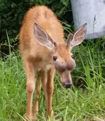
I have seen multiple pictures of deer that look like the one in this image – large, round swellings on the face in front of the eyes. In most of the deer I’ve seen, the swellings have occurred on both sides of the head and been of approximately the same size. And although I’ve received quite a few pictures over the years, very few of these deer have been physically examined.
Based on a very small number of cases, there are 2 rule-outs that lead the pack for this type of swelling.
Chronic sinusitis – otherwise known as inflammation of the sinuses. Bacteria are frequently involved in these instances and contribute to the inflammation. There may be predisposing causes for the sinusitis, such as viral infection, trauma, or tooth root infections, but often the chronic nature of the disease and the bacterial infection make it impossible to identify the original cause. In the few cases I’ve examined, the swellings associated with sinusitis caused cysts (“sacs”) containing air and varying amounts of fluid or puss.
Tumors – A variety of neoplasias (“abnormal growth of cells”) can result in tumors on the face. These masses will be composed of solid tissue that may be hard like bone or softer depending on the type of neoplasia.
Other potential causes that would be less likely would include a hematoma (collection of clotted blood) or possibly a cystic disease of unknown origin, which can occur in young horses.
As with all of our photo diagnoses, the only way to confirm would be to physically exam the deer. In this case, we would examine the masses to determine their composition and where they originated in the body. In addition, we would examine the mass tissue microscopically and perform additional laboratory tests to confirm or rule out causes of the lesion.
Examining these cases provides the basis for understanding what is causing the lesion and what the impacts may be for wild populations, domestic animals, and humans. If you see any deer with this type of lesion, please let us know via Ask the Deer Biologist.
-Dr. Justin Brown, PGC Vet
(and his lovely assistant J.T. Fleegle)
If you would like to receive email alerts of new blog posts, subscribe here.
And Follow us on Twitter @WTDresearch